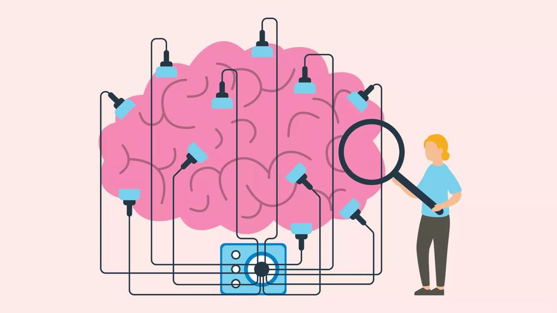
Keeping muscles fit and powerful requires hard work. The same concept holds true for your brain, an amazing organ with the ability to change, adapt and get stronger through mental exercise. ”Neuroplasticity“ is the fancy medical term used to describe your brain’s ability to learn and adapt. Think of it as an internal rewiring process that allows your mind to grow and meet new and increased demands. So, how can you build your brain to flex more mental muscle……….Continue reading….
Source: Cleveland Clininc
.
Critics:
Christopher Shaw and Jill McEachern (eds) in “Toward a theory of Neuroplasticity”, state that there is no all-inclusive theory that overarches different frameworks and systems in the study of neuroplasticity. However, researchers often describe neuroplasticity as “the ability to make adaptive changes related to the structure and function of the nervous system.”[46] Correspondingly, two types of neuroplasticity are often discussed: structural neuroplasticity and functional neuroplasticity.

Structural plasticity is often understood as the brain’s ability to change its neuronal connections. The changes of grey matter proportion or the synaptic strength in the brain are considered as examples of structural neuroplasticity. This type of neuroplasticity often studies the effect of various internal or external stimuli on the brain’s anatomical reorganization. New neurons are constantly produced and integrated into the central nervous system based on this type of neuroplasticity.
Researchers nowadays use multiple cross-sectional imaging methods (i.e. magnetic resonance imaging (MRI), computerized tomography (CT)) to study the structural alterations of the human brains. Structural neuroplasticity is currently investigated more within the field of neuroscience in current academia. Adult neurogenesis “has not been convincingly demonstrated in humans”. Functional plasticity refers to the brain’s ability to alter and adapt the functional properties of network of neurons. It can occur in four known ways namely:
Homologous area adaptation is the assumption of a particular cognitive process by a homologous region in the opposite hemisphere.[49] For instance, through homologous area adaptation a cognitive task is shifted from a damaged part of the brain to its homologous area in opposite side of the brain. Homologous area adaptation is a type of functional neuroplasticity that occur usually in children rather than adults.
In map expansion, cortical maps related to particular cognitive tasks expand due to frequent exposure to stimuli. Map expansion has been proven through experiments performed in relation to the study: experiment on effect of frequent stimulus on functional connectivity of the brain was observed in individuals learning spatial routes.
Cross-model reassignment involves reception of novel input signals to a brain region which has been stripped of its default input. Functional plasticity through compensatory masquerade occurs using different cognitive processes for an already established cognitive task when the initial process cannot be followed due to impairment. Changes in the brain associated with functional neuroplasticity can occur in response to two different types of events:
- previous activity (activity-dependent plasticity) to acquire memory or
- in response to malfunction or damage of neurons (maladaptive plasticity) to compensate a pathological event
In the latter case the functions from one part of the brain transfer to another part of the brain based on the demand to produce recovery of behavioral or physiological processes. Regarding physiological forms of activity-dependent plasticity, those involving synapses are referred to as synaptic plasticity. The strengthening or weakening of synapses that results in an increase or decrease of firing rate of the neurons are called long-term potentiation (LTP) and long-term depression (LTD), respectively, and they are considered as examples of synaptic plasticity that are associated with memory.

The cerebellum is a typical structure with combinations of LTP/LTD and redundancy within the circuitry, allowing plasticity at several sites. More recently it has become clearer that synaptic plasticity can be complemented by another form of activity-dependent plasticity involving the intrinsic excitability of neurons, which is referred to as intrinsic plasticity. This, as opposed to homeostatic plasticity does not necessarily maintain the overall activity of a neuron within a network but contributes to encoding memories.
Also, many studies have indicated functional neuroplasticity in the level of brain networks, where training alters the strength of functional connections. Although a recent study discusses that these observed changes should not directly relate to neuroplasticity, since they may root in the systematic requirement of the brain network for reorganization.
A surprising consequence of neuroplasticity is that the brain activity associated with a given function can be transferred to a different location; this can result from normal experience and also occurs in the process of recovery from brain injury. Neuroplasticity is the fundamental issue that supports the scientific basis for treatment of acquired brain injury with goal-directed experiential therapeutic programs in the context of rehabilitation approaches to the functional consequences of the injury.
Neuroplasticity is gaining popularity as a theory that, at least in part, explains improvements in functional outcomes with physical therapy post-stroke. Rehabilitation techniques that are supported by evidence which suggest cortical reorganization as the mechanism of change include constraint-induced movement therapy, functional electrical stimulation, treadmill training with body-weight support, and virtual reality therapy.
Robot assisted therapy is an emerging technique, which is also hypothesized to work by way of neuroplasticity, though there is currently insufficient evidence to determine the exact mechanisms of change when using this method. One group has developed a treatment that includes increased levels of progesterone injections in brain-injured patients. “Administration of progesterone after traumatic brain injury (TBI) and stroke reduces edema, inflammation, and neuronal cell death, and enhances spatial reference memory and sensory-motor recovery.”
In a clinical trial, a group of severely injured patients had a 60% reduction in mortality after three days of progesterone injections. However, a study published in the New England Journal of Medicine in 2014 detailing the results of a multi-center NIH-funded phase III clinical trial of 882 patients found that treatment of acute traumatic brain injury with the hormone progesterone provides no significant benefit to patients when compared with placebo.
Individuals who have chronic pain experience prolonged pain at sites that may have been previously injured, yet are otherwise currently healthy. This phenomenon is related to neuroplasticity due to a maladaptive reorganization of the nervous system, both peripherally and centrally. During the period of tissue damage, noxious stimuli and inflammation cause an elevation of nociceptive input from the periphery to the central nervous system.

Prolonged nociception from the periphery then elicits a neuroplastic response at the cortical level to change its somatotopic organization for the painful site, inducing central sensitization. For instance, individuals experiencing complex regional pain syndrome demonstrate a diminished cortical somatotopic representation of the hand contralaterally as well as a decreased spacing between the hand and the mouth.
Additionally, chronic pain has been reported to significantly reduce the volume of grey matter in the brain globally, and more specifically at the prefrontal cortex and right thalamus. However, following treatment, these abnormalities in cortical reorganization and grey matter volume are resolved, as well as their symptoms. Similar results have been reported for phantom limb pain.


Leave a Reply